

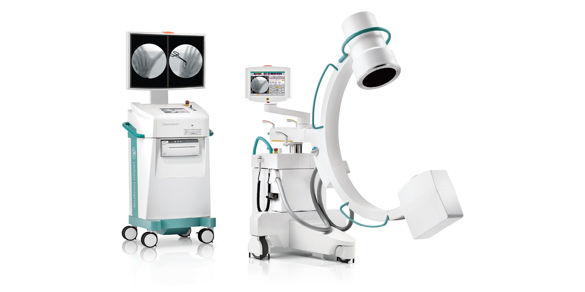
Ziehm Vision² – Call Now for Price
Ziehm Vision² delivers high-quality imaging with low dose exposure on a footprint of just 0.8 m². It features a compact C-arm and monitor cart with an intuitive touchscreen user interface as well as two 19” flatscreen monitors. It has a high-resolution CCD camera that detects over 4,000 shades of gray. The monoblock generator’s unique liquid cooling system (Advanced Active Cooling) is specially designed for extended use in operating theaters, making the Ziehm Vision² ideal for a wide range of clinical applications such as general surgery, orthopedics, and traumatology.
18” TFT monitors provide bright, high contrast image with a wide viewing angle.
Intuitive workflow with synchronized TFT touchscreens on c-arm and monitor cart.
High resolution thanks to a dynamic CCD camera with 1 k × 1 k technology.
Easy integration into existing networks (with WLAN option).
Active Cooling for continuous performance during lengthy, demanding procedures.
Counter-balanced c-arm for simplified positioning.
Ziehm Vision² Specifications
C-Arm Mobile Stand
Dimensions and Mechanics
Motor-driven vertical travel: 17” [43 cm]
Motor-driven horizontal travel: 9” [22 cm]
Motor-driven orbital rotation: -90° / +45°
Angulation: ± 225°
Swiveling (panning): ± 10°
Focus-image receptor distance: 40” [111 cm]
C-arm vertical free space: 35” [89.5 cm]
C-arm depth: 27” [68 cm]
Width: 32″ [80 cm]
Length: 63-72” [160-182 cm]
Height: 62-79” [157.5-200.5 cm]
Brakes: Patented steering & braking lever, with parallel movement of the mobile stand in all directions
X-Ray Generator
X-ray tube: stationary anode
Single focus: 0.6 (IEC 336)
Max. anode heat content: 45 kHU / 34 kJ
Max. anode heat dissipation: 600 W
Generator type:
monoblock
20 kHz high frequency
microprocessor-controlled
Operating Values:
Nominal output (100 kV): 2,000 W
Maximum output: 2,200 W
Pulsed fluoroscopy:
kV range: 40-110 kV
mA range: 0.2-20 mA
pulse width: 10-24 ms
pulse rate: 1, 2, 4, 8, 12.5, 25 pulses/s
Digital radiography (snapshot):
kV range: 40 -110 kV
mA: up to 20 mA
Filtering:
Total filtering: ≥ 4 mm Al equivalent
Output
Pulsed fluoroscopy: up to 20 mA
Digital radiography: up to 20 mA
Tube housing heat capacity:
powered by integrated Advanced Active Cooling and heat management system
5 million HU system heat capacity
400 W continuous heat dissipation in clinical performance
Collimator System
Dedicated pre-collimator for FPD
Collimator rotation: ± 90°
Iris collimator: 2-8” [50-198 mm] diameter
Slot collimator: 2-8” [50-198 mm] diameter
Virtual collimation without radiation
Flat-Panel Detector System (only on FD Model)
Type: amorphous silicon photodiode TFT technology
Scintillator: cesium iodide
Field size: 8 x 8” [19.8 x19.8 cm]
Detector matrix: 1024 x 1024 pixels; 194 μm pixel size
Dynamic range: 72 dB
System resolution: 2.4 lp/mm
Anti-scatter grid: 70 lines/cm; grid ratio 8:1
Laser positioning device integrated in the detector housing
User Interface
TFT touchscreens on C-arm stand and monitor cart: synchronized; intuitive icons for easy use; resolution: 640 x 480 pixels; multi-lingual user interface
Monitor Cart
Monitors
High-resolution and high-brightness twin flat-screen monitors: screen size: 18.1“ (46 cm); native resolution: 1280 x 1024 pixels; viewing angle (horizontal & vertical): 170°; contrast ratio: 600:1; dimensions: 16 x 13 x 3” [41 x 34 x 7 cm]
3D Workstation (for Ziehm Vision2 FD Vario 3D Only)
Hardware:
Intel Core 2
2048 MB DDRAM
USB, CD / DVD (option)
Mouse
Software:
Control software for variable isocentric movement
3D Visualization
Slice planes: axial, saggital, coronal
3D reconstruction algorithm FBP
3D volume size: 5 x 5 x 5” [128 x 128 x 128 mm]
Resolution: 2563 voxel, 5123 voxel
Thick slice filter function
Iso loop
Measurement function: length, angle
Zoom function
Parallel slices function
Digital Image Processing
Real-Time Processing Functions:
Recursive filter: 4 levels (out of 16)
Stack filter (‘Last Image Hold’): 5 levels
Edge enhancement filter: 5 levels
Windowing and step windowing
Digital image rotation and reversal without radiation
Grayscale inversion
Digital shutters
Application-Oriented Anatomical Programs:
Bone, Heart, Abdomen
Soft, Large Patient
Object Detected Dose Control (ODDC):
Automatic motion detection/li>
Automatic metal correction
Image Acquisition:
Autosave
Cine loop with auto-playback (option): sequential image storage and display: 1, 2, 4, 8, 12.5, 25 frames/s; start, stop and replay rate controls
DSA Package (Option):
DSA real-time subtraction with re-masking capability
MSA maximum opacification sequence
RSA roadmapping
Pixel shift / landmarking
Digital measurement functions (distance/angle)
Postprocessing Functions:
Edge enhancement: 5 levels
Zoom: 3 levels
Image rotation
Windowing
Grayscale inversion
Image cropping (digital shutters)
Digital Memory:
Storage capacity: up to 55,000 images; 400 scans ( 2563 voxel volume); 200 scans ( 5123 voxel volume)
Memory matrix: 1024 x 1024 pixels
Image matrix: 1024 x 1024 pixels
Digital image processing: up to 32 bit; 14 bit A/D converter
Grayscale: 1024 shades of gray (10 bit)
Data Organization:
Patient-based data management with 16-image mosaic display
Pre-registration via DICOM Worklist
Manual input or emergency registration
Air Kerma Dose Display
Air Kerma value tagged to stored image


Reviews
There are no reviews yet.